Mole Or Blue Nevus: Is It Dangerous?
Blue nevus is a type of dermatological lesion that appears relatively frequently. It can take different shapes and sizes, but the most characteristic is the intense blue color that differentiates it greatly from other diseases.
Although it is benign, the possibility of malignancy or confusion with a melanoma makes evaluation by a dermatologist highly recommended. Want to know more? Keep reading!
How to identify it?

Although these lesions can appear anywhere on the body, they are most common in the extremities or the sacral coccygeal region. Occasionally, extreme lesions on the head or mucosa may be found that make diagnosis difficult.
From a clinical point of view, the characteristic finding of the blue nevus is its intense color. The shape can encompass several types of primary skin lesions, including macules (small, flat discolorations), papules (here, there is a slight elevation and the content is solid), or nodules, among others.
In either case, the surface has a smooth texture and tends to be less than 1 centimeter, although “giant” lesions may also appear. Some patients can be born with the blue nevus, although most cases are acquired from the third decade of life.
In these latter patients, the moles gradually increase in size over months or years until their growth stops abruptly.
epidemiology
The exact prevalence is unknown. There are approximate measures that state that it affects more Asian people (3 to 5%) than Caucasians (0.5 to 4%), mainly women. Although it can affect all phototypes, it is more common in intermediate skin tone (III and IV according to Fitzpatrick’s classification).
According to other studies, it could occur in 1 in 3,000 live births, although it is an approximate figure that varies depending on the region being considered.
Types of blue nevus
There is still no clear consensus regarding the classification of these injuries. However, in general terms, it is currently possible to distinguish the following forms:
- Common or solitary.
- Mobile.
- Compound.
- Atypical.
- Of large plates.
Despite the clinical differences, this diagnostic classification is applied in the field of histopathology. That is, when a mole biopsy is taken, it is the pathologists who find microscopic changes and make the final diagnosis.
Exceptional cases
Sometimes, it is possible to find moles with unusual clinical and histological characteristics. They generally represent a diagnostic challenge, even during analysis of samples under the microscope.
This case reported in 2009 is about a 7-year-old schoolboy, who was taken to the doctor for presenting repetitive episodes of rhinosinusal inflammation. During the physical examination, turbinate hypertrophy was found that, in part, could explain the pathology.
As part of the routine, an imaging study (computed tomography) was requested to verify the integrity of the paranasal sinuses. In this, important changes were evidenced in the area corresponding to the left maxillary sinus, which under normal conditions should have air.
To determine the origin, a more detailed examination was performed, taking a sample of the lesion, which in fact had a hyperpigmented appearance (dark color). The sample was sent to the pathology laboratory for evaluation, and then a blue nevus was reported. This is an example of a rare presentation of the disease.
Why can it happen?
L os histopathologic findings suggest that blue nevi occur by the presence of melanocytes in the dermis. These are cells capable of producing melanin, the main pigment in the skin. They tend to have strange shapes and are accompanied by changes in the surrounding tissue.
Everything seems to indicate that the cause of the blue nevus would be the combination of genetic and environmental factors. Through clinical research, mutations in G proteins, important molecules that participate in various biochemical reactions called “second messenger signals,” have been identified in nearly every cell in the body.
There is also evidence to suggest that the lesions are the product of embryological defects. The neural crest is a very small primitive structure rich in pluripotent stem cells.
These are capable of “morphing” into various tissues. At a certain point, these cells migrate to various places in the embryo to give rise to the nervous system and other structures, such as melanocytes.
It is likely that some of these cells have migrated in the wrong way, so they end up staying in the dermis and not in the epidermis (which is the most superficial layer of the skin and contains melanocytes). The typical blue coloration of the lesions could be the result of a physical phenomenon called the Tyndall effect.
Blue nevi can occasionally appear in the context of other rare systemic syndromes. Such is the case of the Carney Complex, which is characterized by the appearance of myxomas, changes in skin pigmentation and hormonal problems, such as Cushing’s syndrome.
When is it necessary to go to the doctor?
Although many dermatological lesions are benign and represent no more than a cosmetic discomfort for the patient, this is not always the case. One of the greatest challenges for dermatologists is the early diagnosis and timely treatment of malignant lesions, especially melanoma.
This is the most common that can appear on the skin, and has a great capacity to metastasize and cause damage to other organs. From the pathophysiological point of view, it is characterized by the uncontrolled proliferation of melanocytes, which gives it the characteristic dark color.
Unfortunately, it often goes unnoticed, despite easy-to-identify warning signs. For this, anyone can use the classic ABCDE of melanomas:
- Asymmetry: the lesion does not have a regular shape, so when dividing it into two halves using an imaginary line, differences in size can be found.
- Edges: These tend to be uneven and not follow a well defined line.
- Color: changes in hue are frequent; this is the main reason melanomas might mimic blue nevi.
- Diameter: although they are lesions that can start out very small, they increase in size very quickly and usually exceed 6 millimeters.
- Evolution: They never have a static appearance. As the months go by, they change in any of the aforementioned aspects.
Given the possibility that a simple blue nevus could actually be a malignant melanoma, it is necessary to go to the dermatologist as soon as the lesion is detected.
Diagnosis of blue nevus

When the dermatologist evaluates the lesions, there are two diagnostic phases. The first one is clinical. Through observation, the doctor will establish an initial suspicion. Afterwards, it will be necessary to take a biopsy for the pathologist to make the definitive diagnosis.
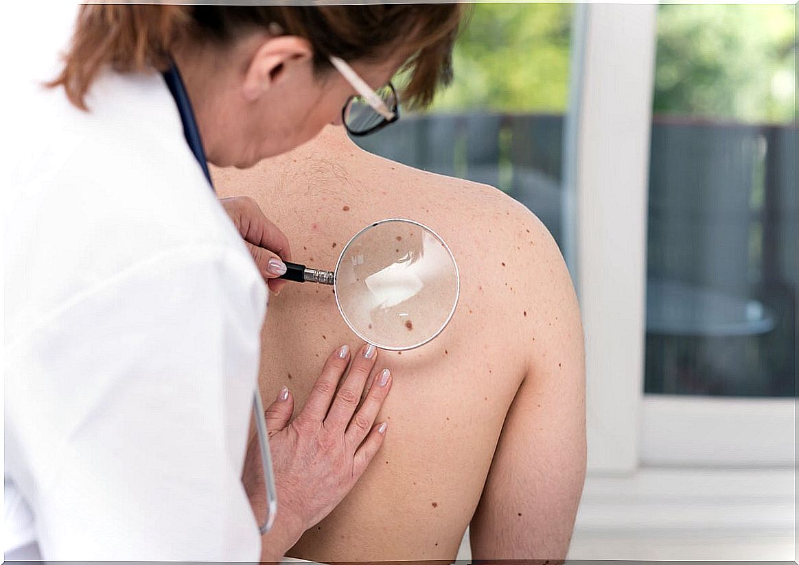
The clinical evaluation usually includes the performance of a dermoscopy, a vital tool for these specialists. It allows you to see the lesions in greater detail by acting as a kind of magnifying glass, requiring an excellent light source to obtain more reliable results.
Thanks to this tool, it is possible to distinguish some characteristics that differentiate the blue nevus from other benign or malignant lesions. This is achieved with different types of lighting, under which moles can reflect a different, easily detectable color.
Histopathologic confirmation is almost always necessary. The main findings were mentioned earlier and, in general, the diagnosis is not difficult to make. Depending on the results, the dermatologist may suggest different treatments.
Differential diagnostics
There are specific varieties of melanoma and other conditions that could be mistaken for blue nevus. Among them it is possible to point out the following
- Dermatofibroma: it is a benign tumor with a hard consistency. It is often confused with a less common form of the blue nevus, known as the amelanocytic form.
- Desmoplastic melanoma : A rare variety that is usually diagnosed only by biopsy.
When is it important to remove it?
If the biopsy determines that it is a benign blue nevus, the lesion may remain untreated. However, periodic medical evaluations are necessary, given the possibility that some subtypes of moles have to become malignant in the future.
In other cases, a more decisive treatment could be chosen, especially when there are aesthetic problems associated with psychological damage. For this, surgical excision is the most appropriate therapy.
The main problem with this treatment modality is that relapses of the disease may appear, in which the doctor may opt for a more conservative attitude. Sometimes the new growths can be accompanied by malignant lesions and, if that is the case, the treatment would be different.
Forecast
In the vast majority of cases, because the lesions are benign, there are no associated complications. In some patients, the lesions can become malignant, so routine medical evaluation with the goal of early treatment is vital.
Fortunately, the blue nevus is benign in most cases. However, in those acquired cases where accelerated growth of the lesions is observed, it is advisable to go to the dermatologist who, together with the pathologist, will establish a definitive diagnosis.


