Contraction Of Skeletal Muscle
Striated muscle is made up of nerves, proteins, motor neurons, blood vessels, and long, cylindrical fibers with more than one nucleus, depending on their need. These fibers are also surrounded by a cell membrane called the sarcolemma.
This muscle is classified as “striated” due to its longitudinal and transverse striations, which maintain the same thickness throughout its entire body, and which are capable of being appreciated through microscopes.
One of the main characteristics of this muscle is that it is of the voluntary type, which is why it develops a rapid contraction and is exhausted.
How many types of skeletal muscle are there?
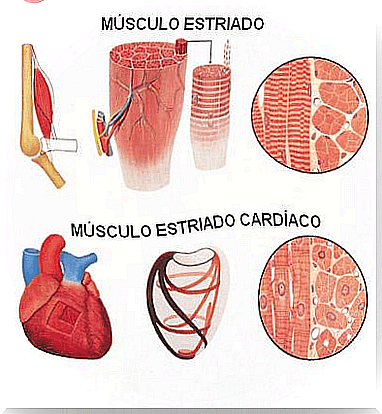
Striated muscle is located throughout the body and can be divided into two types:
- Skeletal. It contains numerous nuclei located on the periphery, near the sarcolemma.
- Cardiac. It contains a single central and unique nucleus, located in the inferior muscle next to the insular vertebra.
Both types present a tonicity, elasticity, contractility and excitability (inserted in the skin or in the skeleton), and have the sarcomere as the fundamental unit for their operation.
What are the characteristics of skeletal muscle?
Skeletal skeletal muscle is of the innervated type. This means that it receives functions from nerve impulses sent by the central nervous system.
Because of this type of functioning under conscious control, it is referred to as a voluntary muscle. Most of these muscles are attached to areas of the skeleton by attachments of connective tissue called the tendon.
The contraction of the muscle allows the movements of the tendons, bones and cartilage of the skeleton, with which, thus, the animals form most of the body.
Unlike skeletal skeletal muscle, cardiac muscle, that is, the heart, is an involuntary muscle. In addition, it is made up of a special striated fiber: the myocardium.
How does contraction work in skeletal muscle?
In the relaxed state, myosin and actin protein fibers overlap each other. This contraction is based on the binding of cytoplasmic calcium ions and troponin covering actin.
On the other hand, this union leaves several points of actin free with myosin, with which there is an increase in cytoplasmic concentrations within the muscle.
For this to be possible, the entry of extracellular calcium through calcium channels is necessary, which produces contraction thanks to the release of the myosin and actin binding sites.
Unlike skeletal skeletal muscle, cardiac muscle maintains its own contraction without the need for motor neurons, so it does not require this process.
And how does relaxation work?
In order to relax, the striated muscle undergoes the breaking of bonds through a process of hydrolysis, thanks to the action of myosin as adenosine triphosphate (ATP). In this process, calcium decreases and makes possible its existence in the sarcoplasmic reticulum that is responsible for storing it.
By decreasing calcium, the cell membrane allows it to escape extracellularly. Eventually, the actin filaments slide into the myosin filaments, thereby allowing skeletal muscle relaxation.
To obtain relaxation, it is necessary to maintain muscle contraction based on adenosine triphosphate (ATP) and disintegrate it with adenosine diphosphate (ADP), in order to provide the required energy.